Mohs Micrographic Surgery
Mohs surgery is a technique used in the treatment of several skin cancers that allows for complete removal of the lesion while minimizing removal of otherwise normal adjacent skin. Any location in the body can be treated with Mohs surgery, but it is typically reserved for nonmelanoma skin cancers occurring on the following locations:
- Ears
- Eyelids
- Nose
- Lips
- Any sensitive location on the body that would have a higher risk of complications with regular surgical excision
Who's At Risk?
In experienced hands, Mohs surgery is a convenient and safe surgical technique that provides precise and complete removal of common nonmelanoma skin cancers while preserving as much normal skin surrounding the lesion as possible. Skin cancers removed typically include:
- Primary basal cell carcinoma
- Primary squamous cell carcinoma
- Recurrent nonmelanoma skin cancers
- Skin cancers with ill-defined borders
- Skin cancers with high recurrence rates
Signs & Symptoms
Depending on physician preference and after careful weighing of risks and benefits, patients on blood-thinning medications may or may not be asked to discontinue these 1–2 weeks prior to the procedure. In most cases, however, patients may safely continue their blood-thinning medications.
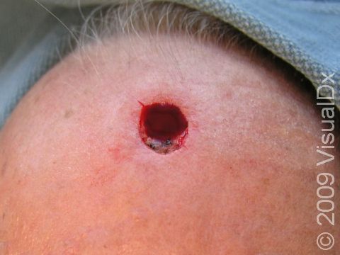
During the procedure, the area is cleaned and prepared for surgery. A needle is used to inject anesthetic into the skin in order to numb the area. A scalpel is used to remove the areas affected with skin cancer plus a very small margin of normal-appearing skin. Each piece that is removed is frozen and then cut into small sections (known as a frozen section), which then allows for microscopic examination done at the time of the surgery. This entire process is called a stage.
If the margins are clear of tumor, the wound is closed. If tumor remains on any one of the frozen sections, it is re-excised, guided by precise mapping techniques that allow for exact localization of the remaining tumor. This is repeated until no more cancer remains. Because one or more stages may be necessary to completely remove the skin cancer, it is a more time-consuming technique compared to a regular surgical excision.
Self-Care Guidelines
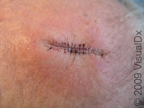
Once the margins of the wound are clear of tumor, the Mohs surgeon then proceeds to repair the wound. The choices of closure technique and repair very much depend on the location and extent of the surgical defect, as well as the experience and preference of the surgeon. Once the defect is closed, a pressure dressing is applied and wound care instructions are discussed with the patient.
The following aftercare is recommended:
- Keep the skin dry during the first 24–48 hours.
- Apply Vaseline® or antimicrobial ointment daily.
- Keep the area covered with gauze dressing or a Band-Aid®.
- Non-dissolving stitches need to be removed in 5–14 days, depending on the type of suture.
Treatments
Each case must be individualized when considering other treatment options. Depending on the type, extent, size, and location of nonmelanoma skin cancers, other treatment options include:
- Electrodesiccation and curettage
- Excision
- Radiation therapy
- Topical creams
- Chemotherapy
When to Seek Medical Care
- Infection
- Bleeding during or after the surgery
- Bruising
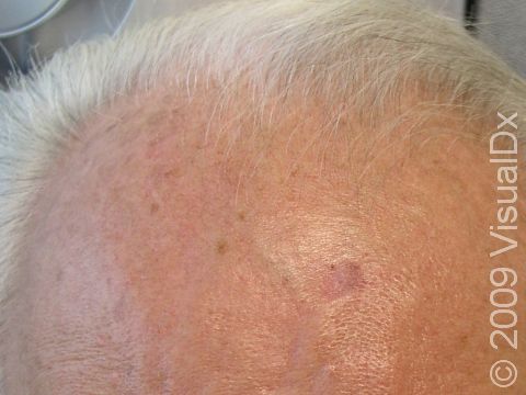
- Scarring
- Wound breakdown
- Contraction of the wound
- Visible stitch marks where the sutures were placed
- Keloid, or a thick (hypertrophic) scar
- There is a risk of cutting important structures such as vessels or nerves, depending on the location and size of the lesion.
Trusted Links
Last modified on October 5th, 2022 at 7:40 pm

Not sure what to look for?
Try our new Rash and Skin Condition Finder


