Milia
Milia are common non-cancerous (benign) skin findings in people of all ages. Milia formed directly from sloughed-off skin, known as primary milia, are small, fluid-filled cysts usually found on the faces of infants and adults, while lesions formed indirectly, known as secondary milia, are small cysts found within areas of skin affected by another skin condition.
Milia are formed when skin does not slough off normally but instead remains trapped in pockets on the surface of the skin. An individual milium is formed from a hair follicle (pilosebaceous unit) or from a sweat gland (eccrine gland). In primary milia in infants, the oil gland (sebaceous gland) may not be fully developed. Secondary milia often develop after injury or blistering of the skin, which disrupts and clogs the tubes (glandular ducts) leading to the skin surface.
Who's At Risk?
Milia can occur in people of all ages, of any race, and of either sex.
Milia are so common in newborn babies (occurring in up to 50% of them) that they are considered normal.
Secondary milia may appear in affected skin of people with the following:
- Blistering injury (trauma) to skin, such as poison ivy
- Burns
- Blistering skin disorders, such as epidermolysis bullosa or porphyria
- Following long-term use of topical steroids
Signs & Symptoms
The most common locations for primary milia include:
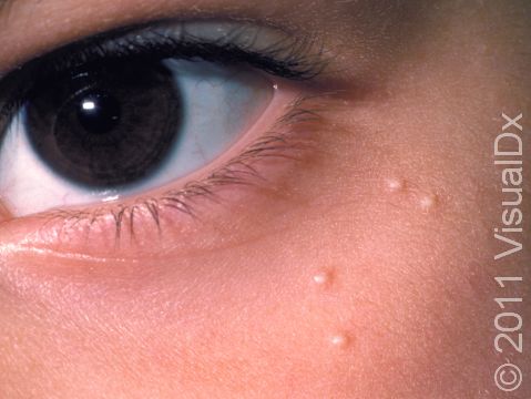
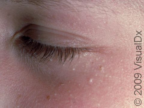
- Around the eye (periorbital area) in children and adults
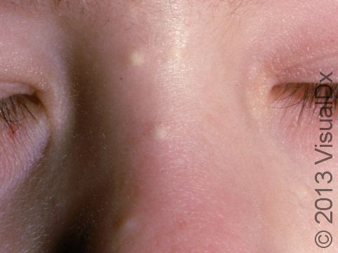
- Around the nose, especially in infants
The most common locations for secondary milia include:
- Anywhere on the body, where another skin condition exists
- On the faces of people who have had a lot of damage from sun exposure
A single lesion (milium) appears as a small (1–2 mm), white-to-yellow, dome-shaped bump on the outer surface of the skin.
Self-Care Guidelines
Although milia are found in the outer (superficial) layers of skin, they are difficult to remove without the proper tools. Do not try to remove them at home as you may leave a scar.
Primary milia found in infants tend to heal on their own within several weeks, though the secondary milia found in older children and adults tend to be long-lasting.
Treatments
If your doctor diagnoses primary milia in an infant, no treatment is necessary as the condition will go away on its own within a few weeks.
If your child has secondary milia, the doctor will likely treat the other skin condition at that area, if it is still present. Other treatments for milia include:
- Topical retinoid cream such as tretinoin, tazarotene, or adapalene
- Removal with a sterile blade (lancet) or scalpel followed by use of a special tool called a comedone extractor
- A series of fruit acid peels or microdermabrasion procedures at the dermatologist’s office
Visit Urgency
See your child’s doctor or a dermatologist for evaluation if you notice any new bump on your child’s skin.
Trusted Links
References
Bolognia, Jean L., ed. Dermatology, pp.1722-1723. New York: Mosby, 2003.
Freedberg, Irwin M., ed. Fitzpatrick’s Dermatology in General Medicine. 6th ed. pp.601, 604, 780. New York: McGraw-Hill, 2003.
Last modified on August 16th, 2022 at 2:45 pm

Not sure what to look for?
Try our new Rash and Skin Condition Finder


