Kerion
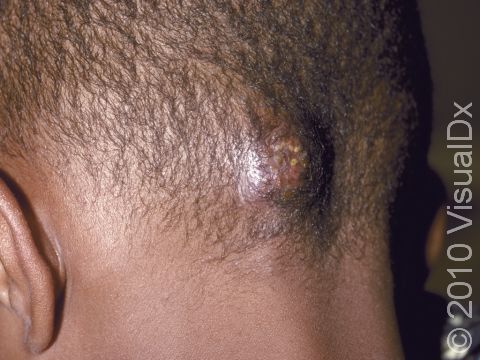
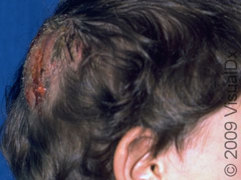
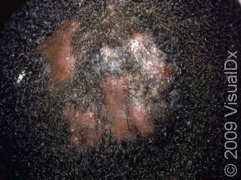
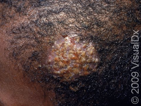
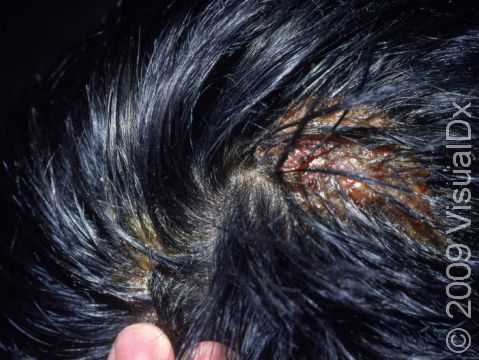
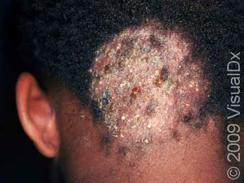
A kerion is a scalp condition that occurs in severe cases of scalp ringworm (tinea capitis). A kerion appears as an inflamed, thickened, pus-filled area, and it is sometimes accompanied by a fever.
The underlying condition, scalp ringworm, is a usually harmless fungal infection of the scalp and hair that occurs as scaly spots and patches of broken hair on the head. Ringworm of the scalp is most commonly seen in children. Though several different species of fungus may cause scalp ringworm, they are generally known as dermatophytes. Scalp ringworm may be acquired by direct contact with infected people or with contaminated objects that have been handled by infected people (such as combs, pillows, and sofas). Most commonly, scalp ringworm infections are caused by dermatophytes that prefer to grow on humans. Less commonly, the fungus may be spread from infected animals (zoophilic dermatophytes) or from the soil (geophilic dermatophytes).
Kerions usually occur in people who have been infected with zoophilic dermatophytes. A kerion is believed to be an overly active response of the immune system or an allergic reaction to the fungus.
Who's At Risk?
Scalp ringworm may occur in people of all ages, of all races, and of both sexes. However, it occurs most commonly in children.
A kerion is seen almost exclusively in children, but, on rare occasions, it may be seen in teens and young adults.
Signs & Symptoms
A kerion appears as a thick, mushy area of the scalp. Its surface is often studded with pus-filled bumps (pustules). The kerion can break open and drain pus. If untreated, a kerion can lead to scarring and permanent hair loss (alopecia).
Fever and pain may accompany the kerion. In addition, the lymph nodes at the back of the scalp, behind the ears, or along the sides of the neck may be swollen.
Self-Care Guidelines
There are no effective self-care measures to treat a kerion.
Treatments
Often, the doctor is able to diagnose a kerion just by looking at it. However, in order to confirm the diagnosis, the physician may wish to scrape some surface skin scales onto a slide and examine them under a microscope. This procedure, called a KOH (potassium hydroxide) preparation, allows the doctor to look for tell-tale signs of fungal infection.
Sometimes the doctor will also perform a fungal culture in order to document the presence of fungus or to discover the particular organism that is causing the kerion. The procedure involves:
- Plucking a few hairs or piercing any pus-filled lesions in the involved areas of the scalp
- Rubbing a sterile cotton-tipped applicator across the skin to collect scale and pus
- Sending the specimen away to a laboratory
Typically, the laboratory will have results within 2–3 weeks. In some cases, the laboratory is able to identify the particular type of dermatophyte that is causing the scalp ringworm and kerion. The doctor may also perform a bacterial culture in the same manner to see if there is a bacterial infection present.
Occasionally, a Wood’s lamp is used to look for the fungus. In this procedure, the doctor shines a black light at the scalp, and certain strains of dermatophytes may appear as yellow-green fluorescent spots when seen under this light.
A kerion is treated with oral antifungal medicines because the fungus grows deep into the hair follicle where topical creams and lotions cannot penetrate. Scalp ringworm and kerion usually require at least 6–8 weeks of treatment with oral antifungal pills or syrup, including:
- Griseofulvin
- Terbinafine
- Itraconazole
- Fluconazole
- Ketoconazole
Often, the doctor will also prescribe a medicated shampoo to reduce the risk of spreading the infection to someone else:
- Selenium sulfide shampoo
- Ketoconazole shampoo
If the bacterial culture is positive (shows bacterial growth), the physician may want to start your child on an oral antibiotic as well.
If the kerion is particularly tender and painful, your child’s doctor may recommend starting oral corticosteroids (cortisone pills or syrup). Steroids are strong medications that can quickly reduce the inflammation present in the kerion.
Visit Urgency
See your child’s doctor for evaluation if your child loses hair or has itchy, scaly spots on the scalp. If your child develops a thick, pus-filled pocket on the scalp, see the doctor soon to evaluate for kerion.
Trusted Links
References
Bolognia, Jean L., ed. Dermatology, pp.1179-1180. New York: Mosby, 2003.
Freedberg, Irwin M., ed. Fitzpatrick’s Dermatology in General Medicine. 6th ed. pp.1861, 1994. New York: McGraw-Hill, 2003.
Last modified on October 5th, 2022 at 7:36 pm
Not sure what to look for?
Try our new Rash and Skin Condition Finder
