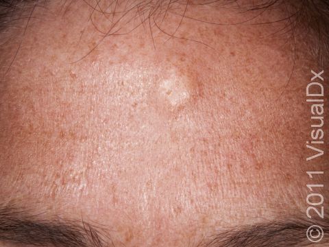
Epidermoid cysts are lined by skin cells that form a scale that accumulates in the cyst and appears as a creamy substance.
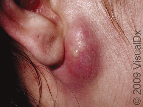
This image displays a large, infected cyst, which is draining some pus.
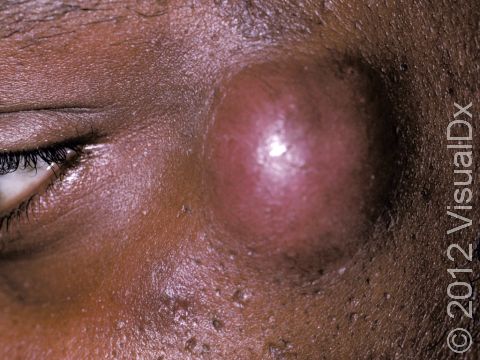
This image displays a large, tense cyst at the temple.
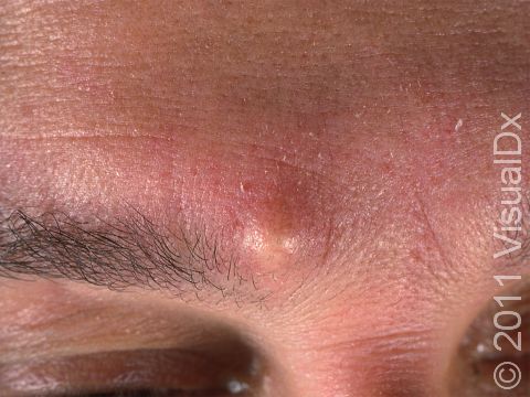
An epidermoid cyst can appear as a visible lump, and the patient may complain that it feels much larger than it looks.
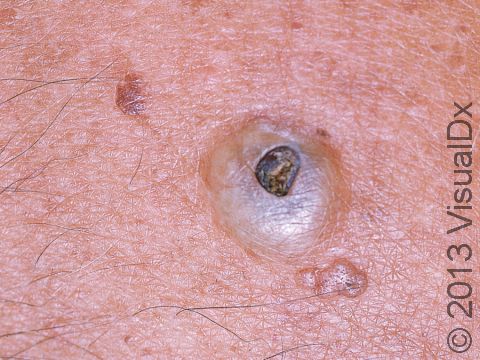
This image displays a cyst that has opened to the surface over time, turning a dark brown and appearing as a
This image displays a cyst that has opened to the surface over time, turning a dark brown and appearing as a "plug" through the opening.
With careful examination, a dilated hair follicle can be seen in the middle of an epidermoid cyst. It often goes unnoticed until the cyst becomes infected and pus drains from the follicle.

Images of Epidermoid Cyst (6)

Epidermoid cysts are lined by skin cells that form a scale that accumulates in the cyst and appears as a creamy substance.
This image displays a large, infected cyst, which is draining some pus.
This image displays a large, tense cyst at the temple.
An epidermoid cyst can appear as a visible lump, and the patient may complain that it feels much larger than it looks.
This image displays a cyst that has opened to the surface over time, turning a dark brown and appearing as a
With careful examination, a dilated hair follicle can be seen in the middle of an epidermoid cyst. It often goes unnoticed until the cyst becomes infected and pus drains from the follicle.

Epidermoid Cyst

Epidermoid cysts, sometimes known as sebaceous cysts (a misnomer), contain a soft “cheesy” material composed of keratin, a protein component of skin, hair, and nails.

  • Epidermoid cysts form when the top layer of skin (epidermis) grows into the middle layer of the skin (dermis). This may occur due to injury or blocked hair follicles.
  • The lesion may be asymptomatic, but rupture of the epidermoid cyst can result in significant discomfort.


Who's At Risk?

Epidermoid cysts are a common lesion that affect people of all ages.

Signs & Symptoms

Epidermoid cysts can be located almost anywhere but are most common on the face, neck, scalp, or trunk.

  • A cyst appears as a dome-shaped, skin-colored growth that usually moves when touched and pressed upon. It may have a small opening in the center.
  • The cyst can be well-defined or irregular due to prior rupture, scarring, and regrowth.
  • If manipulated or infected, the cyst can become red and may be tender.

Self-Care Guidelines

None necessary. It is advised not to try to express the material within cysts as further inflammation and even infection may result.

Treatments

  • Inflamed, non-infected cysts may be injected with steroids to reduce inflammation.
  • Incision and drainage can provide immediate reduction in the cyst. However, this is a temporary measure. After this treatment, a cyst will refill with the cheesy contents because the lining of the cyst has not been removed.
  • Cysts may be removed (excised) surgically.

Visit Urgency

See your primary care physician or a dermatologist if a cyst becomes inflamed or painful.

References

Bolognia, Jean L., ed. Dermatology, pp.1721-1723. New York: Mosby, 2003.

Freedberg, Irwin M., ed. Fitzpatrick’s Dermatology in General Medicine. 6th ed, pp.778-781. New York: McGraw-Hill, 2003.

Last modified on October 5th, 2022 at 7:22 pm
Not sure what to look for?

Try our new Rash and Skin Condition Finder
Age
Gender
Submit